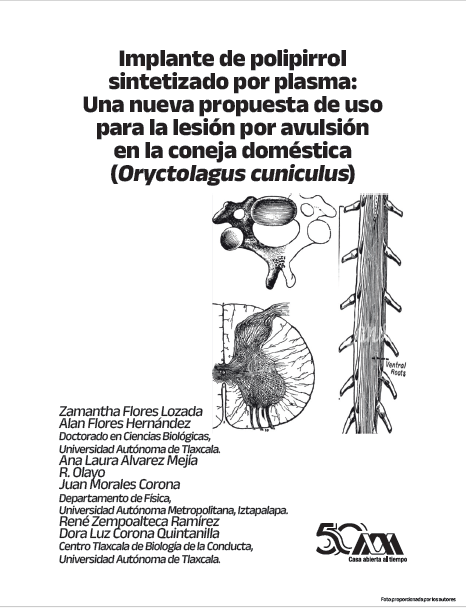
Implante de polipirrol sintetizado por plasma
Una nueva propuesta de uso para la lesión por avulsión en la coneja doméstica (Oryctolagus cuniculus)
Abstract
Locomotion is a fundamental behavior that allows animals to move in different environments and directions. Movement is regulated by sensorimotor circuits at the level of the spinal cord, specifically in the lumbar segments. One of the injuries that affect these segments is the separation of the spinal cord from the nerve roots. If the separation occurs in the ventral horn of the spinal cord, it is called a ventral root avulsion (VRA). Due to the complex pathophysiology of VRA, it is necessary to development efficient strategies to repair and regenerate the avulsed nerve fibers. In recent years, the use of implants created from polymeric materials has gained relevance in the biological and medical field. Polypyrrole is one of the most studied polymers and, due to its electroconductive properties, is a biomaterial of interest for axonal regeneration. This has led to the proposal of using polypyrrole implants as a treatment for axonal damage caused by avulsion.
Downloads
References
Corona-Quintanilla, Dora Luz, Acosta-Ortega, C., Flores-Lozada, Z., López-Juárez, R., Zempoalteca, R., Castelán, F., & Martínez- Gómez, M. (2020). Lumbosacral ventral root avulsion alters reflex activation of bladder, urethra, and perineal muscles during micturition in female rabbits. J Neurourol Urodyn, 39(5), pp. 1283–1291.
Cruz GJ, Mondragón-Lozano R, Diaz- Ruiz A, Manjarrez J, Olayo R, Salgado-Ceballos H, et al (2012). Plasma polypyrrole implants recover motor function in rats after spinal cord transection. J Mater Sci Mater Med, 23(10), pp. 2583-92.
Deliagina, T. G., Zelenin, P. V., Beloozerova, I. N., & Orlovsky, G. N. (2007). Nervous mechanisms controlling body posture. Physiol Behav, 92(1–2), pp. 148–154.
Eggers, Ruben, Tannemaat, M. R., De Winter, F., Malessy, M. J. A., & Verhaagen, J. (2016). Clinical and neurobiological advances in promoting regeneration of the ventral root avulsion lesion. Eur J Neurosci, 43(10), pp. 318–335.
Farley, A., Johnstone, C., Hendry, C., & McLafferty, E. (2014). Nervous system: part 1. Nurs Stand, 28(31), pp. 46–51.
Flores-Lozada Z. (2021). Efecto de la avulsión unilateral de L6 sobre la conducción nerviosa a nivel de raíz dorsal de cinco nervios periféricos de la extremidad inferior en la coneja (Oryctolagus cuniculus) [Tesis de maestría, Universidad Autónoma de Tlaxcala]. Repositorio Institucional de la Universidad Autónoma de Tlaxcala https://repositorio.uatx.mx:8443/bitstream/ DSyTI_UATx/631/1/Flores%20Lozada% 20Zamantha.pdf
Hejcl, A., Urdzikova, L., Sedy, J., Lesny, P., Pradny, M., Michalek , J., Burian , M., Hajek , M., Zamecnik, J., Jendelova, P., & Sykova, E. (2008). Acute and delayed implantation of positively charged 2-hydroxyethyl methacrylate scaffolds in spinal cord injury in the rat. J Neurosurg Spine, 8(1), pp. 67-73.
Olayo R, Alvarez-Mejía AL, Mondragon-Lozano R, Escalona A, Morales C, Morales I, et al. Implante de polímeros sintetizados por plasma en lesión de médula. In: garcíacolín L, Dagdug L, Picquart, editors. La Física biología en México: Temas selectos 2: Colegio Nacional, pp. 195 – 205, 2008.
Pérez-Estudillo C. A, Sánchez-Alonso D, López-Meraz, M. L., Morgado-Valle C, Beltran-Parrazal, L., Coria-Avila G. A., Hernández Aguilar M. E, Manzo-Denes, J. (2018). Aplicaciones terapéuticas para la lesión de médula espinal. eNeurobiología, 9(21), pp. 1–16.


